
Early Warning Signs of Heart Failure You Shouldn’t Ignore
Heart failure announces itself quietly, in pillows, bathroom scales, and abandoned stairs. The early warning signs
Ultrasound imaging of the heart — showing how well it pumps, how the valves are working, and the size and thickness of each chamber.
An echocardiogram is an ultrasound scan of the heart. Gel is applied to the chest and a probe is moved across it, producing moving images of the heart as it beats.
It is painless, involves no radiation, and takes around thirty minutes. It is the single most informative non-invasive test in cardiology, because it shows structure and function at the same time.
Where an EKG shows the electrical activity, the echocardiogram shows the mechanics — how forcefully the heart squeezes, how well it relaxes, and whether the valves open and close properly.
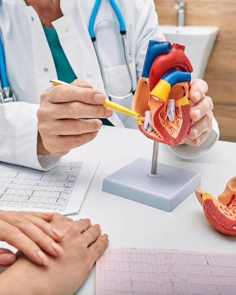



An echocardiogram produces a great deal of data, and its value lies in the interpretation. Our cardiologists review your study themselves and explain what the measurements mean for you rather than handing over a report.

General & Preventive Cardiology

Preventive & Non-Invasive Cardiology

Interventional & Vascular Cardiology
Treatment plans tailored to your symptoms, history, and individual needs.
Preventive, diagnostic, interventional, and vascular care in one practice.
Modern diagnostics for heart rhythm, function, structure, and blood flow.
Serving Pearland with additional locations expanding across Greater Houston.
Experienced physicians with advanced cardiovascular training and expertise.
We work with many leading insurance plans for convenient access to care.
Alliance Heart & Vascular is expanding to serve patients across Pearland, Greater Houston and surrounding communities.
Comprehensive cardiology, preventive cardiovascular care, diagnostic evaluation, and follow-up.
View Pearland LocationAlliance Heart & Vascular is expanding across Greater Houston and nearby communities, bringing convenient access to trusted cardiovascular care closer to where our patients live and work.
Alliance Heart & Vascular works with many major health insurance plans. Insurance participation can vary by plan, network, employer, and location.
Please call (346) 614-0002 before your appointment to verify your specific insurance benefits and network participation. Our team can help answer questions about scheduling, referrals, and insurance verification.
An echocardiogram is arranged whenever the question concerns how the heart is working rather than how it is beating. Common reasons include:
If heart failure is suspected, an echocardiogram is essential rather than optional. It is the test that separates the two main types, and they are treated differently.
Echocardiography is central to diagnosing several cardiovascular conditions. Learn more about:
An echocardiogram is a scheduled test. If you have severe chest pain, sudden breathlessness, or fainting, call 911 immediately.
Do not wait for an office appointment.
Online appointment requests should never be used for potentially life-threatening symptoms.
A single scan produces a large amount of information about the structure and performance of the heart:
Ejection fraction is the number most patients are told, but it is only part of the picture. A heart with a normal ejection fraction can still be stiff and failing to fill properly.
Each part of the study answers a different clinical question, and together they build a complete picture of how your heart is performing.
Ejection fraction measures the percentage of blood the left ventricle ejects with each beat. It defines which type of heart failure a patient has.
Each of the four valves is assessed for narrowing and leakage, and the severity is graded to determine whether monitoring or treatment is needed.
Enlargement or thickening reveals the effect of long-standing high blood pressure, valve disease, or cardiomyopathy.
How well the heart relaxes and fills between beats. Impairment here causes heart failure even when the pumping percentage looks normal.
Segments that move poorly can indicate previous heart attack or an area of muscle with a reduced blood supply.
Fluid around the heart, and estimated pressures in the lungs and right side, which matter in breathlessness and swelling.
A standard echocardiogram requires no special preparation at all. To make the appointment straightforward:
You may hear whooshing sounds during the scan. That is blood flow being measured with Doppler, and it is a normal part of the study rather than a sign of a problem.
The echocardiogram is the definitive non-invasive test for several conditions, and a major contributor to the assessment of many more.
The echocardiogram measures ejection fraction and filling, which is what separates reduced from preserved heart failure. Read more about heart failure.
It distinguishes a cardiac cause of breathlessness from a pulmonary one, which symptoms alone cannot do. Read more about shortness of breath.
Thickening of the heart muscle from long-standing high blood pressure is identified and measured. Read more about high blood pressure.
Areas of muscle moving poorly can indicate previous damage or a reduced blood supply. Read more about coronary artery disease.
An echocardiogram at diagnosis assesses chamber size, valve disease, and pumping function, all of which guide treatment. Read more about atrial fibrillation.
Not every patient needs every cardiac test. Testing should be selected based on your symptoms, examination, medical history, and cardiovascular risk. Depending on your individual situation, your cardiologist may recommend one or more of the following.
Measures the heart's electrical activity and may identify rhythm problems or other cardiac changes.
Uses ultrasound to evaluate heart function, chambers, valves, muscle, and structure.
Shows how the heart responds to physical activity and may help evaluate exercise-related symptoms.
Uses cardiac imaging during stress testing to assess blood flow to the heart muscle.
Records heart rhythm over time to help detect intermittent arrhythmias or palpitations.
Blood tests may evaluate cholesterol, blood sugar, kidney function, and other cardiovascular risk factors.
Certain patients may require additional cardiac imaging or coronary evaluation depending on their symptoms and risk profile. Your cardiologist will explain why a particular test is being recommended and how the results may influence your treatment plan.
The study is reviewed by your cardiologist and explained in the context of your symptoms. What follows depends on the findings and may include:
Valve disease and heart failure are both conditions where the interval between scans matters. Your cardiologist will tell you when the study should be repeated and why.
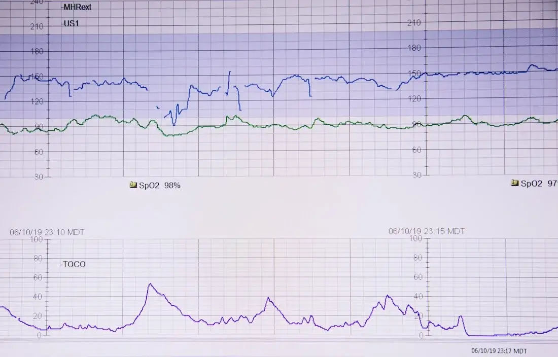
Murmurs are commonly picked up at routine examinations, and reduced pumping function is sometimes found incidentally during a hospital admission. Both need proper assessment: many murmurs are entirely innocent, while others indicate valve disease that should be tracked.
An appointment is worthwhile if you have:
Talk to our office — Monday to Friday, 8:00 a.m. to 4:30 p.m.
Call (346) 614-0002One scan shows both structure and function.
Whether you have been referred for an echocardiogram, have a murmur that needs assessing, or have breathlessness or swelling without a clear explanation, we perform and interpret echocardiograms on site at both Houston locations.
New patients are welcome.
Serving patients throughout Pearland, Greater Houston, and surrounding areas.